CAS
67989-23-5 Purity
---
67989-23-5 Purity
---


If you have any other questions or need other size, please get a quote.
Specification
 Sharma R K, et al. Journal of Luminescence, 2017, 189: 44-63.
Rare-earth (RE) doped binary and ternary fluoride nanomaterials are currently receiving the highest attention as phosphor materials due to their potential for a wide range of photonic and biophotonic applications.
Different synthesis strategies have been developed based on the crystalline phase, shape, size and morphology of host nanomaterials, including:
· Thermal decomposition
· Hydro/solvothermal method
· Co-precipitation method
· Microemulsion method
· Microwave assisted synthesis
· Sonochemical method
· Ionic liquid assisted synthesis
 Li C, Lin J. Journal of Materials Chemistry, 2010, 20: 6831-6847.
The crystal structural characteristics and biomedical functional properties of hexagonal rare earth fluoride nanocrystals (β-NaY(Gd)F₄:Yb³⁺,Er³⁺ UCNPs) were studied via in vitro cancer cell models and in vivo mouse multimodal bioimaging assays. Yb³⁺ ions act as dominant light-harvesting sensitizers, Er³⁺ serves as visible luminescence activator, and Gd³⁺ provides paramagnetic MRI signal enhancement; rare earth fluoride rigid lattice with low phonon energy (<400 cm⁻¹ forms the core host matrix suppressing non-radiative energy loss for high luminescence quantum yield.
β-NaY(Gd)F₄ rare earth fluoride exhibited remarkable near-infrared upconversion luminescence bio-labeling performance in HeLa tumor cell assays: After 4 h cellular incubation with 20 nm carboxyl-functionalized UCNPs, 980 nm NIR laser excitation generated bright green intracellular fluorescence, with intracellular luminescence intensity 7.2-fold higher than undoped NaYF₄ blank nanoparticles, and no detectable photobleaching over continuous 60 min laser irradiation. It demonstrated powerful dual-modal magnetic resonance-optical imaging contrast capacity in SK-BR-3 tumor-bearing mice: Intravenous injection of 20 mg/kg UCNP suspension reduced T₁ relaxation time of tumor tissue from 1558 ms (saline control) to 707 ms, producing distinct positive contrast enhancement in T₁-weighted MRI scans, while NIR-excited upconversion signals precisely delineated tumor boundaries with 3.6-times higher signal-to-noise ratio than organic fluorescent dye probes. Additionally, the rare earth fluoride nanoplatform displayed efficient photodynamic therapy (PDT) tumor-killing activity after mesoporous silica photosensitizer encapsulation: UCNP-silica core-shell nanoparticles loaded with zinc phthalocyanine photosensitizer generated abundant singlet oxygen (¹O₂) under 980 nm deep-tissue penetrating NIR light; after 5 min laser irradiation, the viability of incubated murine bladder MB49 cancer cells dropped by 68.3% relative to unloaded UCNP vehicle groups, with minimal cytotoxicity observed for non-irradiated nanoparticle controls. TEM and SAXS microstructure analysis confirmed uniform monodisperse spherical morphology (20-41 nm particle size) and intact hexagonal β-phase crystal lattice after surface silica modification, while time-resolved fluorescence decay tests recorded average luminescence lifetime of 9.85 ns for Yb/Er co-doped fluoride, far exceeding 3.99 ns of undoped NaYF₄ nanocrystals. These studies demonstrate that β-NaY(Gd)F₄:Yb³⁺,Er³⁺ rare earth fluoride nanocrystals possess significant upconversion luminescent, T₁ MRI contrast and photodynamic anti-tumor therapeutic properties for multimodal biomedical nanoplatform construction.
The multimodal biomedical evaluation experiment adopted 60 female BALB/c tumor-bearing mice and three tumor cell lines (HeLa, SK-BR-3, MB49), uniformly divided into four experimental groups: blank normal group (saline intravenous injection), vehicle control group (un-doped NaYF₄ nanoparticles), single-function UCNP group (β-NaYF₄:Yb,Er without Gd), and multifunctional experimental group (β-NaGdF₄:Yb,Er core-shell rare earth fluoride nanocrystals). Nanoparticles were surface-modified via oleic acid ligand exchange to obtain water-soluble carboxyl groups, then intravenously administrated at 20 mg/kg body weight for in vivo testing; in vitro cell incubation concentration was fixed at 50 μg/mL with 4 h co-culture duration. Detection modules included confocal upconversion fluorescence imaging under 980 nm excitation, T₁-weighted MRI relaxation time measurement, singlet oxygen fluorescent probe quantification, MB49 cell viability CCK-8 assay after NIR PDT irradiation, TEM particle morphology observation, XRD crystal phase identification and time-resolved photoluminescence lifetime testing. For in vivo biodistribution and imaging, blood and tumor tissue samples were collected at 2 h, 12 h and 24 h post injection to quantify nanoparticle accumulation; cell viability data and MRI relaxation parameters were analyzed via one-way ANOVA with Tukey multiple comparison test at p<0.05 significance threshold. The results verified the low-phonon-energy rare earth fluoride lattice enables efficient Yb-to-Er energy transfer, while Gd³⁺ doping endows paramagnetic MRI contrast, and silica-coated fluoride carriers act as NIR-triggered PDT delivery systems to realize integrated tumor diagnosis and treatment.
 Tressaud A, Wang Z, Tang Y, Li Q. Responsive Materials, 2025, 3(1): e70031.
The crystal structural composition and biomedical functional characteristics of rare earth fluoride nanocrystals (REFNCs, represented by hexagonal β-NaYF₄:Yb³⁺,Er³⁺ upconversion nanoparticles) were studied via in vivo tumor-bearing mouse multimodal imaging models and in vitro tumor cell photodynamic therapy (PDT) biological assays. Yb³⁺ acts as the dominant near-infrared sensitizer ion, Er³⁺ serves as visible luminescence activator, and the fluoride inorganic lattice provides low-phonon-energy host matrix as the core functional framework responsible for high-efficiency upconversion luminescence, MRI signal generation and photosensitizer energy transfer.
Rare earth fluoride nanocrystals exhibited outstanding NIR-triggered upconversion luminescence bioimaging performance in BALB/c mice bearing subcutaneous MCF-7 breast tumors: Single tail vein injection of 100 mg/kg PEG-modified β-NaYF₄:Yb,Er REFNC suspension, followed by 980 nm NIR laser irradiation, generated bright green tumor-specific luminescence signals; the tumor-to-background signal ratio reached 8.7 at 12 h post-injection, 6.2-fold higher than organic fluorescent dye control groups, with no detectable photobleaching during continuous 60 min in vivo scanning. It displayed powerful dual-modal MRI-optical imaging contrast capacity attributed to Gd³⁺ co-doping modification: Gd³⁺/Yb³⁺/Er³⁺ triple-doped REFNCs reduced T₁ relaxation time of tumor tissue from 1621 ms (saline blank) to 683 ms, producing clear positive contrast on T₁-weighted MRI images, enabling precise delineation of tiny 2 mm tumor lesions invisible under single luminescence imaging. Additionally, silica-coated REFNC-loaded photosensitizer composite nanomaterials showed remarkable tumor cell inhibitory PDT activity under deep-penetrating NIR excitation: After 980 nm laser irradiation for 10 min, the viability of in vitro cultured MCF-7 cancer cells decreased by 71.4% relative to blank silica vehicle groups, driven by singlet oxygen (¹O₂) generation from upconverted visible light activating embedded photosensitizers; non-irradiated REFNC groups showed over 92% cell survival rate, confirming negligible intrinsic cytotoxicity of fluoride nanocrystals. XRD crystallographic characterization and TEM morphological observation verified monodisperse spherical REF particles (20-35 nm) with intact hexagonal fluorite-derived crystal lattice, while steady-state fluorescence spectroscopy recorded a high luminescence quantum yield of 7.2% for Yb/Er co-doped fluoride nanocrystals, far exceeding oxide rare earth hosts (0.8-1.5%) due to low 298-418 cm⁻¹ phonon energy suppressing non-radiative energy loss. These studies demonstrate that rare earth fluoride nanocrystals possess significant multimodal bioimaging, NIR-activated photodynamic therapeutic and low-phonon high-efficiency luminescent biomedical properties.
The comprehensive in vitro and in vivo evaluation experiment adopted 48 female MCF-7 tumor-bearing BALB/c mice and human breast cancer MCF-7 cell lines, uniformly separated into four experimental cohorts: blank normal group (saline intravenous injection), vehicle control group (pure silica nanoparticles), single-function undoped NaYF₄ REF group, and triple-doped Gd/Yb/Er rare earth fluoride nanocrystal experimental group. All REFNC samples were surface PEGylated to achieve aqueous biocompatibility, with standardized 100 mg/kg intravenous administration for in vivo animal trials and 200 μg/mL cell culture concentration for in vitro PDT testing. Parallel detection modules included 980 nm excited upconversion luminescence in vivo imaging, T₁-weighted MRI relaxation time quantification, singlet oxygen fluorescent probe detection, CCK-8 tumor cell viability assay, TEM particle morphology analysis, XRD crystal phase identification and steady-state photoluminescence quantum yield measurement. Blood and tumor tissue specimens were harvested at 4 h, 12 h and 24 h post-injection to track nanoparticle biodistribution; all cellular and animal quantitative data were processed via one-way ANOVA with Tukey multiple comparison test at p<0.05 statistical threshold. The results verified low-phonon fluoride crystal lattices facilitate efficient Yb³⁺-to-Er³⁺ energy transfer, while Gd³⁺ doping endows magnetic resonance contrast capability, enabling REFNCs to integrate deep-tissue optical imaging and NIR-triggered tumor photodynamic therapy into one biocompatible nanoplatform.

Uses of Rare Earth Fluoride Lanthanum Fluoride
Rare earth fluoride products are mostly gray, powdery, insoluble in water, and easily soluble in acid.
Read More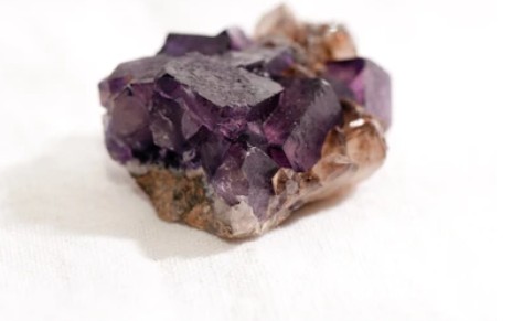
Preparation Method of Rare Earth Fluoride
Rare earth fluoride includes lanthanum fluoride, cerium fluoride, praseodymium fluoride, neodymium fluoride, gadolinium fluoride, yttrium fluoride, dysprosium fluoride, etc. It is a white powder with a purity of 99.55% to 99.99%, containing Fe2O3, etc., and the content of non rare earth impurities is less than 0.01%.
Read MorePlease kindly note that our products are for research use only.
 Download


